Kyphosis
Quasimodo, the Hunchback of Notre Dame, is a figure of our collective imagination. But did you know that the character actually suffered from a spinal deformity called kyphosis or hyperkyphosis?
Hyperkyphosis
The spine, seen from the sagittal plane (profile view), has four physiological curves: the cervical lordotic curve (nape of the neck), thoracic kyphotic curve (upper back between the shoulder blades), lumbar lordotic curve (dip in the lower back) and the sacrococcygeal kyphotic curve (coccyx).
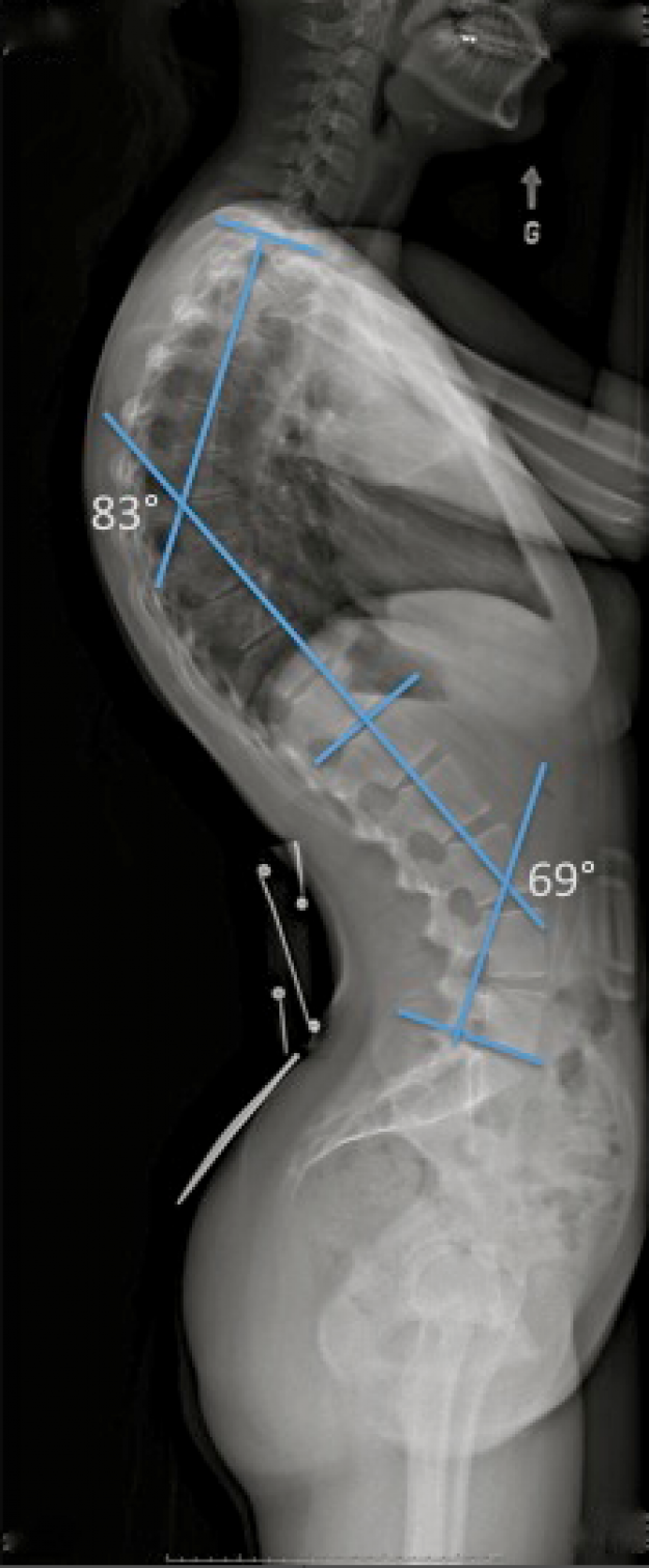
Hyperkyphosis describes an increase in the normal thoracic kyphotic curve. It also often referred to as kyphosis.
 |
 |
|
X-ray of hyperkyphosis |
Hyperkyphosis |
 |
 |
|
|
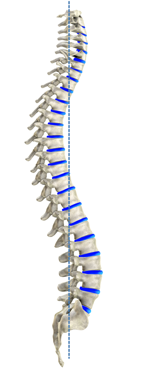
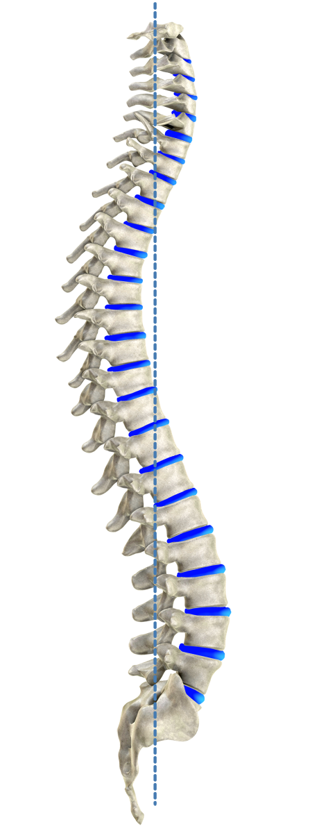
Ideal spinal model by Harrison |
||
There are several types of kyphosis:
- Childhood or adolescent kyphosis, although often blamed on bad posture habits, is actually most frequently due to abnormal vertebral growth, as in Scheuermann's disease.
- Kyphosis in young adults is most commonly related to inflammatory diseases of the spine, or spondylarthropathies.
- Acquired lumbar kyphosis in adults, also known as camptocormia or bent spine syndrome (BSS), is often caused by vertebral compression (osteoporosis), multiple degenerative disc diseases (aging of the intervertebral discs), neurological deficit or atrophy of paravertebral muscles.
Kyphosis can very often be painful.
Morphological classification of kyphosis
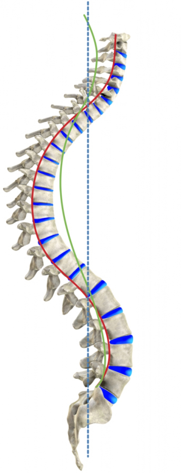
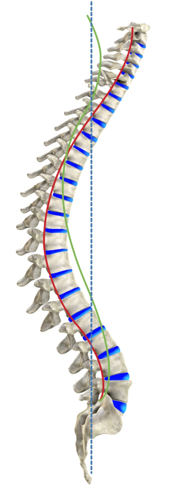
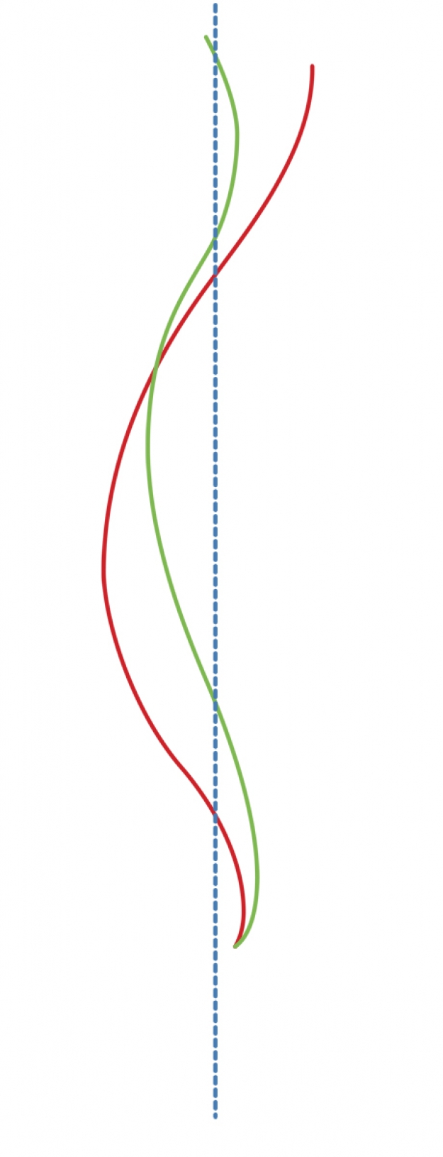
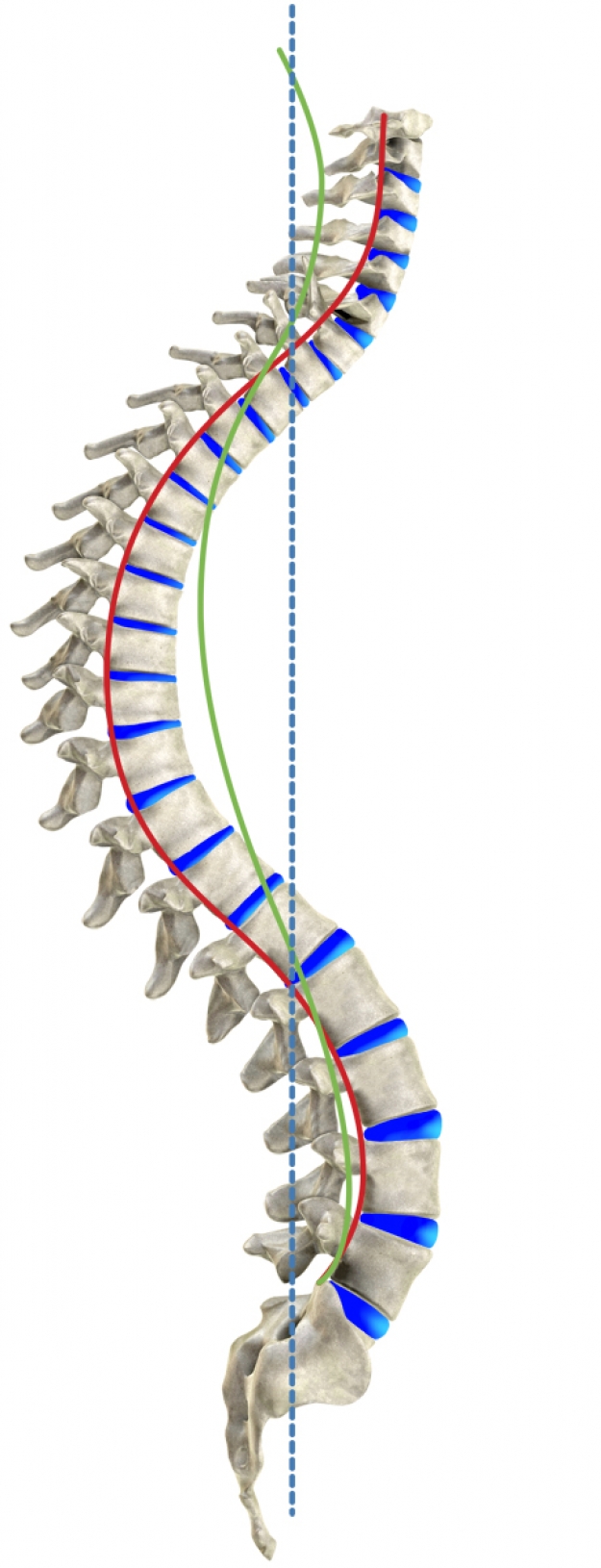
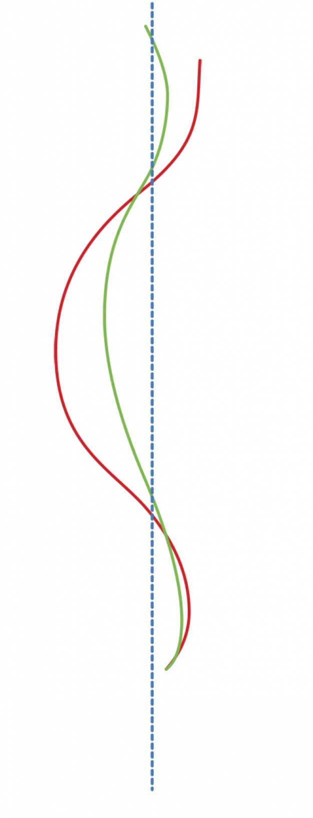
In 2009, chiropractor Dr. Louise Marcotte, B.Sc. D.C. proposed a morphological classification of kyphosis, describing the three basic forms, usually associated with a forward projection of the head (46):
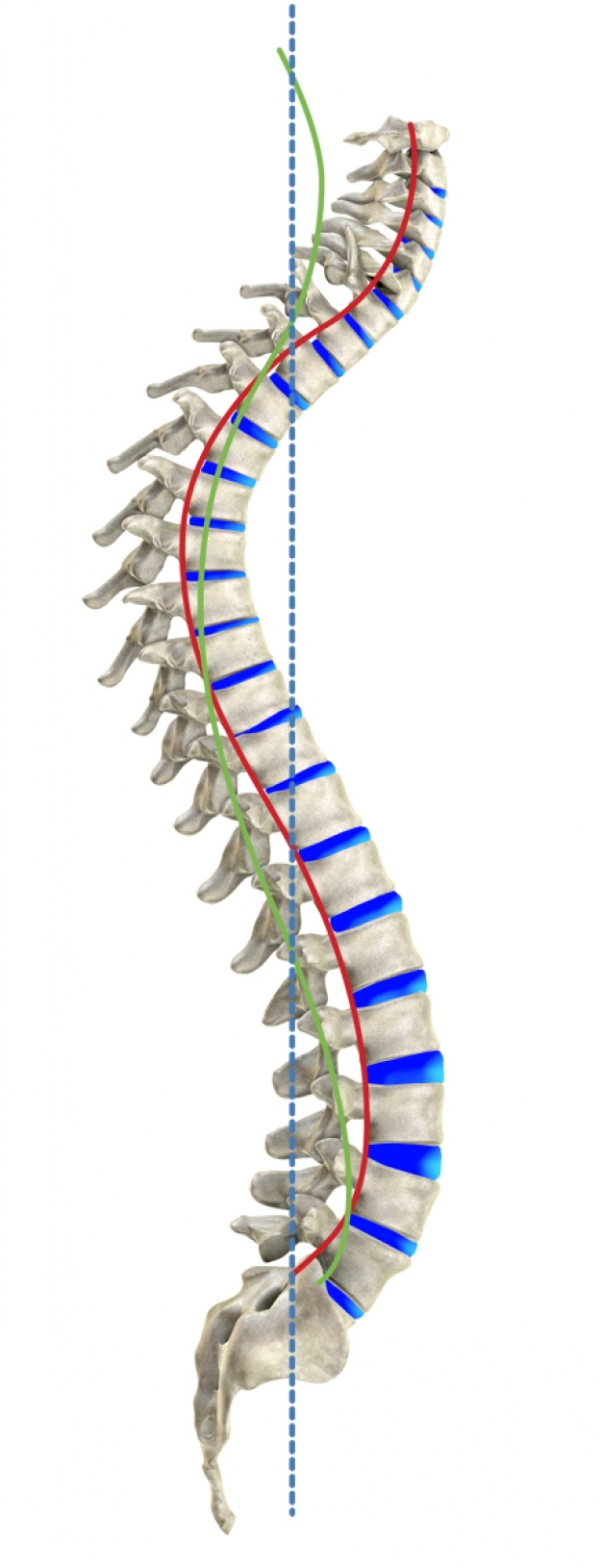
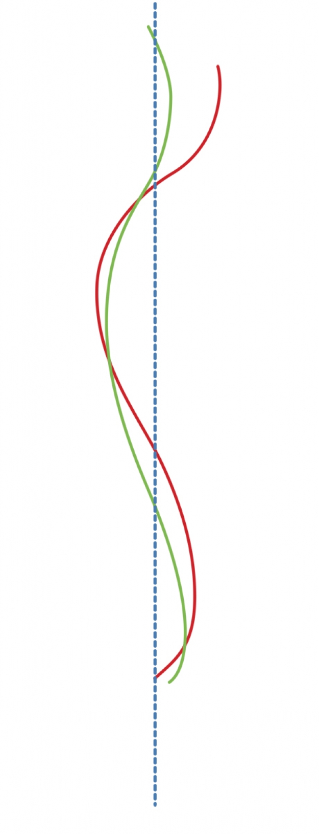
Note: The green lines represent the ideal spinal curves as established by Harrison's ideal spinal model. The red lines represent the curves of a spine affected by kyphosis.
  |
  |
|
Ideal spinal model by Harrison |
Type I hyperkyphosis (low): The apex of the kyphotic deformity is located between the T9 and L1 vertebrae and is most often associated with posterior translation of the thoracic cage relative to the pelvis, as well as flexion of the thoracic cage. |
  |
  |
|
Ideal spinal model by Harrison |
Type II hyperkyphosis (middle): The apex of the kyphotic deformity is usually located between the T7 and T10 vertebrae and is not usually associated with anterior or posterior translation of the thoracic cage relative to the pelvis, but rather with extension of the thoracic cage and the higher lumbar vertebrae. |
  |
  |
|
Ideal spinal model by Harrison |
Type III hyperkyphosis (high): The apex of the kyphotic deformity is usually located between the T5 and T7 vertebrae and is frequently associated with anterior translation of the thoracic cage relative to the pelvis, as well as with extension of the thoracic cage. |
 |
 |
 |
 |
 |
 |
|
Type I |
Type I |
Type II |
Type II |
Type III |
Type III |
This morphological classification enables medical professionals to develop an effective corrective movement strategy to treat hyperkyphosis using the SpineCor® dynamic brace and/or OrthoCyphology chiropractic methods.
To see how the SpineCor® brace can treat kyphosis, visit the SPINECOR section.


